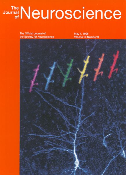
Time-lapse confocal imaging of neuronal dendrites in live, developing hippo-campal tissue slices revealed rapid and dramatic changes in the structure of postsynaptic spine-like protrusions. The illustration depicts a fluorescently labeled CA1 pyramidal neuron (white cell) with developing dendrites in a live tissue slice derived from a neonatal rat. The higher-magnification color images, progressing in a time sequence from left to right (5 min intervals), reveal changes in the structure of individual spiny protrusions along a segment of dendrite from the cell shown. Such dynamic changes in dendritic structure during development may contribute to the formation and plasticity of synaptic contacts with axons.
See: Michael E. Dailey and Stephen J Smith. "The dynamics of dendritic structure in developing hippocampal slices". Journal of Neuroscience. 1996 May 1;16(9):2983-94.