Microglia
GFP+ Microglia & Sytox Orange-labeled nuclei in P4 mouse hippocampus.
( CX3CR1GFP/+)
Images by L. Fuller and M. Dailey.
Image A
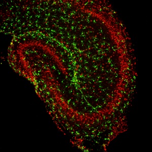
Image B
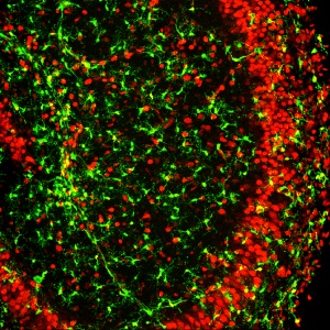
Image C
Microglia & YFP+ Neurons in P12 mouse hippocampus
GFP+ Microglia & YFP+ Neurons in P12 mouse hippocampus
(CX3CR1GFP/+:Thy1-YFP)
Images by L. Fuller and M. Dailey.
Image 1
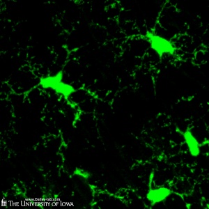
Image 4
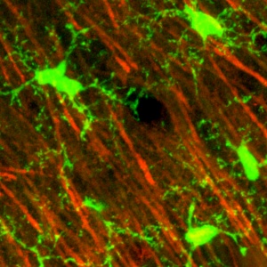
Image 2
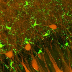
Image 5
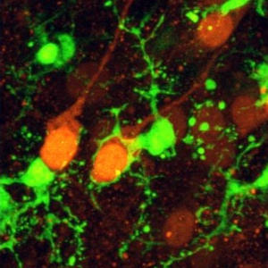
Image 3
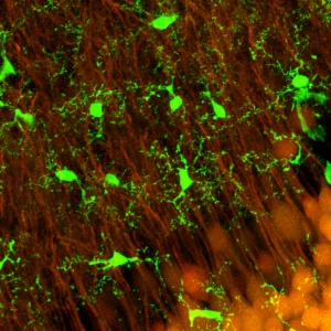
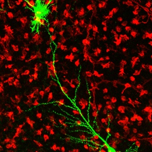
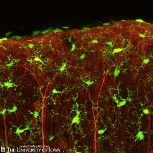
GFP+ Microglia & YFP+ Neurons in P12 mouse neocortex
Image 1
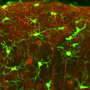
Image 4
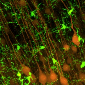
Image 2
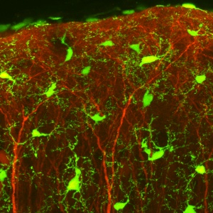
Image 3
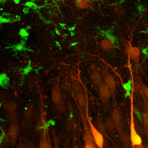
Neurons and activated microglia in mouse hippocampal slice culture in vitro
Neurons and activated microglia in mouse hippocampal slice culture in vitro
(CX3CR1GFP/+:Thy1-YFP)
Images by L. Fuller and M. Dailey.
Image 1
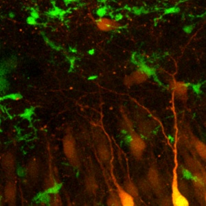
Image 2
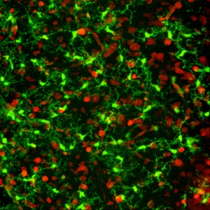
Activated microglia, astrocyte, and pyramidal neuron in rat hippocampal slice culture
Activated microglia, astrocyte, and pyramidal neuron in rat hippocampal slice culture.
All microglia in the field of view are labeled by IB4-lectin (red). Only one neuron and one astroctye are expressing GFP introduced by gene gun-mediated transfection.
Images by D. Kurpius, R. Ahmed, and M. Dailey.
Image 1
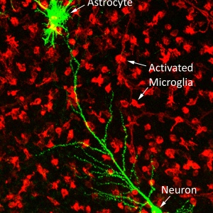
Image 2