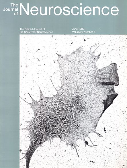
A whole-mount electron micrograph of a growth cone from a rat superior cervical ganglion neuron culture prepared by direct freezing from the living state followed by freeze substitution and critical-point drying. The living growth cones can be stained with a fluorescent dye, DiOC6, which allows the visualization of the dynamics of the endoplasmic reticulum and other membranous organelles.
See: Michael E. Dailey and Paul C. Bridgman. "Dynamics of the endoplasmic reticulum and other membranous organelles in growth cones of cultured neurons". Journal of Neuroscience. 1989 Jun;9(6):1897-909.