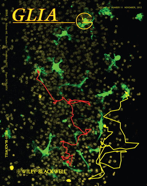
Dual-channel confocal microscope image showing migration tracks (yellow and red lines) of two microglial cells (green) around the CA1 neuronal cell body layer in an acutely isolated postnatal day 2 (P2) mouse hippocampal slice subjected to oxygen and glucose deprivation (OGD). Dots indicate starting points of migration, and arrowheads indicate ending points after 6hr. The mobility and viability of microglia during OGD is developmentally regulated.
See: Eyo & Dailey, "Effects of oxygen-glucose deprivation on microglial mobility and viability in developing mouse hippocampal tissues." Glia. 2012 60(11):1747-1760.
Image by Ukpong Eyo.