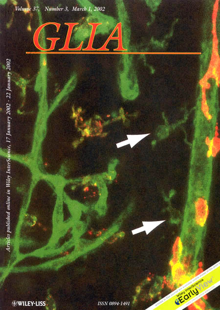
Confocal image of double-labeled rat hippocampal tissue showing microglial and perivascular cells in relation to brain microvessels. Parenchymal microglia and microvessels were labeled with a fluorescently conjugated isolectin, FITC-IB4 (green). Perivascular cells, which affiliate with arterioles and venules, were labeled with antibodies against the ED-2 antigen (red). Perivascular parenchymal microglia (arrows) are ED-2 negative.
See: Grossmann R, Stence N, Carr J, Fuller L, Waite M, and Dailey ME. "Juxtavascular microglia migrate along brain microvessels following activation during early postnatal development." Glia. 2002 Mar 1;37(3):229-40.
Image by Jenny Carr.